定位你的能量小體MitoSpy? Mitochondrial Probes

免疫熒光(IF)的應用
MitoSpy? Orange和 MitoSpy? Red定位于線粒體是基于膜電位,因此MitoSpy? Orange和 MitoSpy? Red能很好的反應細胞的健康狀態和定位。MitoSpy?Orange CMTMRos和MitoSpy?Red CMXRos含有一個氯甲基團,它能共價與細胞中的半胱氨酸連接,這使得細胞在經過固定破膜后,仍能保持在細胞中。但由于固定破膜過程中洗掉了大部分的試劑,所以這需要使用更高的試劑濃度來獲得充分的染色(具體濃度可參考下表)。
而MitoSpy? Green FM不同于之前的兩個,它定位于線粒體不是基于膜電位。它不含有氯甲基,而是含有氟甲基,所以固定破膜之后,大部分的MitoSpy? Green會被洗掉。所以需要固定破膜的樣本,不建議使用該試劑。
|
細胞條件 |
MitoSpy?Orange推薦濃度 |
MitoSpy?Red推薦濃度 |
MitoSpy? Green推薦濃度 |
|
活細胞 |
50-250nM |
50-250nM |
50-250nM |
|
固定的細胞 |
50-250nM |
50-250nM |
50-250nM |
|
固定破膜后的細胞 |
250-500nM |
250-500nM |
不推薦 |
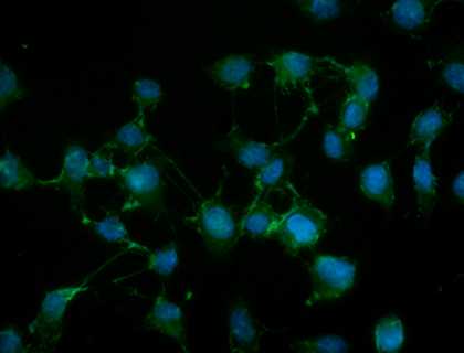
NIH3T3 cells were stained with 100 nM of MitoSpy? Red CMXRos (red) for 20 minutes at 37°C, fixed with 1% paraformaldehyde (PFA) for ten minutes at room temperature, and permeabilized with 1X True Nuclear? Perm Buffer for ten minutes at room temperature. Then the cells were stained with Flash Phalloidin? NIR 647 (green) for 20 minutes at room temperature and counterstained with DAPI (blue). The image was captured with a 60x objective
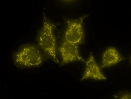
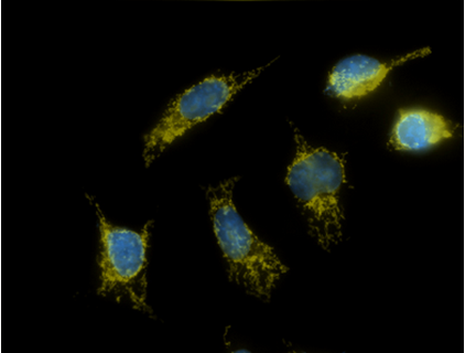
Live staining (50 nM) Fix/Perm staining(500nM)
MitoSpy? Orange
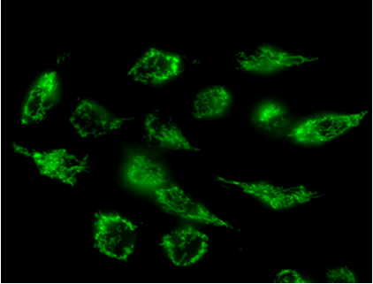
HeLa cells that were stained live with either MitoSpy? Orange (yellow) or Green (green). Cells that were fixed and permeabilized with 4% PFA and 0.1% Triton X-100 were also stained with DAPI (blue). Photos were taken with a 60x magnification.
流式應用(FC)
MitoSpy?Orange CMTMRos和MitoSpy?Red CMXRos也可作為細胞健康的指標。當線粒體活躍的呼吸時,線粒體膜之間存在潛在的差異,稱為膜極化。如果細胞正經歷凋亡或死亡,則該探針不會被該細胞的線粒體強烈吸引。如下圖所示,MitoSpy?Orange和MitoSpy?Red的陽性細胞群是活細胞和健康的細胞,而Annexin V陽性細胞群是凋亡的早期階段的細胞。在流式應用中,這兩種試劑不應在分析前被固定,因為固定會有大量的試劑流失。如果試劑經過固定后丟失,信號強度的損失會混淆確定表型凋亡的能力。所以 MitoSpy?Orange和MitoSpy?Red的固定僅適用于成像應用中的亞細胞定位。
而對于MitoSpy? Green,但現在MitoSpy? Green FM的完整機制還不是很清楚,可能與該試劑的結構和線粒體表面的蛋白之間的同源性相關。由于該試劑的膜電位的獨立性,可以用于流式中測量單個細胞的線粒體量。
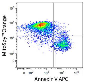
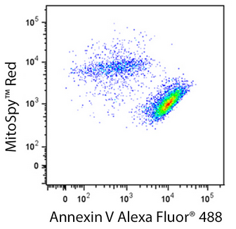
Human T-cell leukemia cell line, Jurkat, was treated for 5 hours with LEAF? purified anti-CD95 (clone EOS9.1), then stained with an impermeant nucleic acid stain, APC or Alexa Fluor? 488 Annexin V, and either MitoSpy? Orange or MitoSpy? Red as indicated. Nucleic acid stain positive events were excluded from analysis.
|
|
|
|
|
|
適合細胞類型 |
適合樣本 |
應用 |
固定 |
|||
|
Cat# |
產品名稱 |
Ex/Em |
等效通道 |
亞細胞定位 |
活細胞 |
死/固定細胞 |
組織 |
細胞 |
流式 |
顯微鏡 |
用PFA處理* |
|
424805/ 424806 |
MitoSpy? Green FM |
488 nm/520 nm |
FITC |
線粒體 |
● |
|
|
● |
● |
● |
|
|
424803/ 424804 |
MitoSpy? Orange CMTMRos |
551 nm/576 nm |
Alexa Flour?555,PE |
線粒體 |
● |
|
|
● |
● |
● |
● |
|
424801/ 424802 |
MitoSpy? Red CMXRos |
577 nm/598 nm |
Alexa Flour?594 |
線粒體 |
● |
|
|
● |
● |
● |
● |


